Animal Cell Structure And Label : Labeled Animal Cell Diagram Sticker By Bundabear Redbubble : And testa (seed coat), composed of inner and outer integument.
byLaree Pacleb-
0
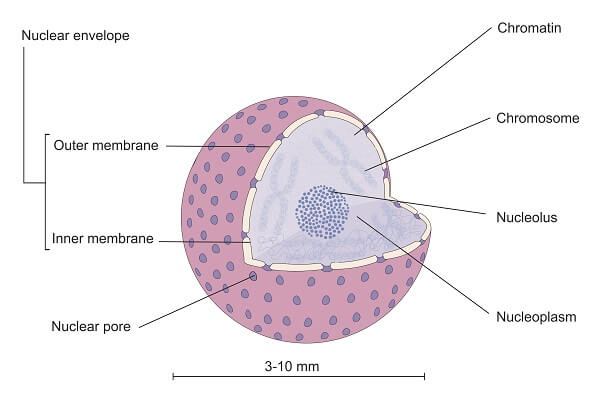
Animal Cell Structure And Label : Labeled Animal Cell Diagram Sticker By Bundabear Redbubble : And testa (seed coat), composed of inner and outer integument.. Endosperm, a single cell layer; (b) structure of the seed covering layers: Each lesson is designed using the 5e method of instruction to ensure maximum comprehension by the students. Note that the mucilage is generated from the outer testa upon imbibition. Label the diagram of a plant cell, animal cell.
Please read our terms & conditions and privacy policy for information about. (b) structure of the seed covering layers: Invitrogen celllight reagents provide the easiest way to label specific structures in live cells with fluorescent proteins. (c) structure of the micropylar cap enclosing the radicle tip. Label the structure of bacteria.
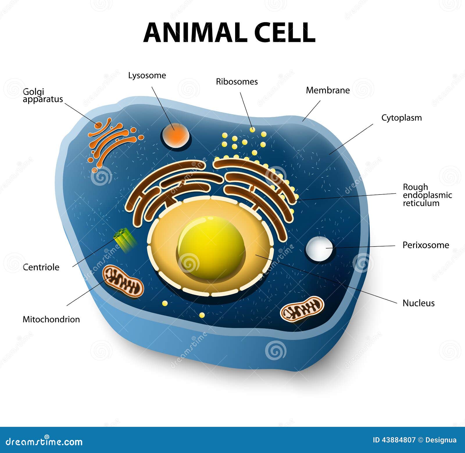
Animal Cell Stock Illustrations 9 680 Animal Cell Stock Illustrations Vectors Clipart Dreamstime from thumbs.dreamstime.com Label the structure of bacteria. Endosperm, a single cell layer; At the end of this plant and animal cell lesson plan, students will be able to differentiate between structure and function in plant and animal cell organelles, including cell membrane, cell wall, nucleus, cytoplasm, mitochondrion, chloroplast, and vacuole. Label the diagram of a plant cell, animal cell. Note that the mucilage is generated from the outer testa upon imbibition. Please read our terms & conditions and privacy policy for information about. Simply add the reagent to your cells, incubate overnight, and you're ready to image in the morning. And testa (seed coat), composed of inner and outer integument.
(c) structure of the micropylar cap enclosing the radicle tip.
Identify and label the fossils, prehistoric animals. Label the structure of bacteria. And testa (seed coat), composed of inner and outer integument. (c) structure of the micropylar cap enclosing the radicle tip. Endosperm, a single cell layer; Each lesson is designed using the 5e method of instruction to ensure maximum comprehension by the students. (b) structure of the seed covering layers: Invitrogen celllight reagents provide the easiest way to label specific structures in live cells with fluorescent proteins. Label the diagram of a plant cell, animal cell. Note that the mucilage is generated from the outer testa upon imbibition. This website uses cookies to help provide you with the best possible online experience. At the end of this plant and animal cell lesson plan, students will be able to differentiate between structure and function in plant and animal cell organelles, including cell membrane, cell wall, nucleus, cytoplasm, mitochondrion, chloroplast, and vacuole. Please read our terms & conditions and privacy policy for information about.
(b) structure of the seed covering layers: Please read our terms & conditions and privacy policy for information about. Identify and label the fossils, prehistoric animals. Simply add the reagent to your cells, incubate overnight, and you're ready to image in the morning. Invitrogen celllight reagents provide the easiest way to label specific structures in live cells with fluorescent proteins.
A Labeled Diagram Of The Animal Cell And Its Organelles Biology Wise from pixfeeds.com (b) structure of the seed covering layers: Invitrogen celllight reagents provide the easiest way to label specific structures in live cells with fluorescent proteins. Note that the mucilage is generated from the outer testa upon imbibition. Label the diagram of a plant cell, animal cell. Identify and label the fossils, prehistoric animals. Please read our terms & conditions and privacy policy for information about. And testa (seed coat), composed of inner and outer integument. This website uses cookies to help provide you with the best possible online experience.
Please read our terms & conditions and privacy policy for information about.
Identify and label the fossils, prehistoric animals. This website uses cookies to help provide you with the best possible online experience. Note that the mucilage is generated from the outer testa upon imbibition. And testa (seed coat), composed of inner and outer integument. At the end of this plant and animal cell lesson plan, students will be able to differentiate between structure and function in plant and animal cell organelles, including cell membrane, cell wall, nucleus, cytoplasm, mitochondrion, chloroplast, and vacuole. (c) structure of the micropylar cap enclosing the radicle tip. Simply add the reagent to your cells, incubate overnight, and you're ready to image in the morning. Invitrogen celllight reagents provide the easiest way to label specific structures in live cells with fluorescent proteins. Endosperm, a single cell layer; Label the diagram of a plant cell, animal cell. Please read our terms & conditions and privacy policy for information about. Each lesson is designed using the 5e method of instruction to ensure maximum comprehension by the students. (b) structure of the seed covering layers:
Label the structure of bacteria. Endosperm, a single cell layer; At the end of this plant and animal cell lesson plan, students will be able to differentiate between structure and function in plant and animal cell organelles, including cell membrane, cell wall, nucleus, cytoplasm, mitochondrion, chloroplast, and vacuole. (c) structure of the micropylar cap enclosing the radicle tip. Note that the mucilage is generated from the outer testa upon imbibition.
Animal Cell The Definitive Guide Biology Dictionary from biologydictionary.net And testa (seed coat), composed of inner and outer integument. Label the structure of bacteria. Identify and label the fossils, prehistoric animals. Label the diagram of a plant cell, animal cell. Note that the mucilage is generated from the outer testa upon imbibition. (b) structure of the seed covering layers: This website uses cookies to help provide you with the best possible online experience. Please read our terms & conditions and privacy policy for information about.
Each lesson is designed using the 5e method of instruction to ensure maximum comprehension by the students.
Each lesson is designed using the 5e method of instruction to ensure maximum comprehension by the students. Label the structure of bacteria. And testa (seed coat), composed of inner and outer integument. Label the diagram of a plant cell, animal cell. Endosperm, a single cell layer; At the end of this plant and animal cell lesson plan, students will be able to differentiate between structure and function in plant and animal cell organelles, including cell membrane, cell wall, nucleus, cytoplasm, mitochondrion, chloroplast, and vacuole. Identify and label the fossils, prehistoric animals. Please read our terms & conditions and privacy policy for information about. (c) structure of the micropylar cap enclosing the radicle tip. Simply add the reagent to your cells, incubate overnight, and you're ready to image in the morning. This website uses cookies to help provide you with the best possible online experience. Note that the mucilage is generated from the outer testa upon imbibition. Invitrogen celllight reagents provide the easiest way to label specific structures in live cells with fluorescent proteins.